CASE NOTES
First reported case of ovine-herpesvirus-2-associated systemic vasculitis and mortality in an Australian Wiltshire Horn flock
Babu K Nath2,3, Mitchell L5, Tridip das1, Pankaj Dhar1, Renate Schwab2, Jade K Forwood 2,3, Shane R Raidal4 and Shubhagata Das1,3
- School of Agricultural, Environmental and Veterinary Sciences, Faculty of Science and Health, Charles Sturt University, Wagga Wagga, New South Wales 2678, Australia
- Biosecurity Research Program and Training Centre, Gulbali Institute, Charles Sturt University, Wagga Wagga, New South Wales 2678, Australia
- Training Hub Promoting Regional Industry and Innovation in Virology and Epidemiology, Gulbali Institute, Charles Sturt University, Wagga Wagga, New South Wales 2678, Australia
- Melbourne Veterinary School, Faculty of Science, The University of Melbourne, 250 Princes Highway, Werribee, VIC, 3030, Australia
- Previously from Forbes Veterinary Clinic
Posted Flock and Herd June 2026
INTRODUCTION
Domestic and wild sheep are recognised as asymptomatic natural reservoirs for ovine herpesvirus 2 (OvHV-2), the causative agent of sheep-associated malignant catarrhal fever (SA-MCF) in susceptible species such as cattle, deer, and bison. Although sheep are considered well-adapted hosts in which infection is typically subclinical, recent reports have described molecular and histopathological evidence of MCF-like systemic necrotising vasculitis associated with OvHV-2 infection in sheep (Phillips et al., 2018; Sheehan et al., 2024). Here, we describe a case of clinical disease and death associated with OvHV-2 infection in a nine-month-old Wiltshire Horn ewe lamb. To the best of our knowledge, this case represents the first documented report of such a disease presentation in sheep in Australia.
CASE DESCRIPTION
On 25 May 2023, a nine-month-old Wiltshire Horn ewe lamb presented for examination at a private veterinary clinic with a history of "nose bleeds" and worsening lethargy. She was in moderately good condition (BCS 2.5/5) and weighed 48 kg. Physical examination revealed mild, diffuse opacity of the right eye and pale mucous membranes. She was mildly febrile; however, as she had been transported to the clinic, this finding was attributed to stress. The remainder of the physical examination was unremarkable, and she was reported to be eating, drinking, urinating, and defaecating normally. History provided by the owner indicated that she had experienced three episodes of unilateral epistaxis from the left nostril on 12, 19, and 22 May.
The flock was kept on unimproved native pastures, with water provided in troughs from a rainwater tank. Feed supplementation was conducted on an as-needed basis and generally consisted of good-quality lucerne hay. The sheep were provided access to salt and mineral blocks. Lambs were vaccinated with Glanvac 6 (Zoetis) at marking (eight weeks of age), with a booster given five weeks later. Gudair (Zoetis) vaccination was administered at approximately 10 weeks of age. Adult ewes were generally drenched with a drench containing multiple actives in June/July based on faecal egg counts. This ewe lamb had been recently drenched with Zolvix Plus (25 mg/mL monepantel, 2 mg/mL abamectin; Elanco).
The ewe did not respond to treatment with cloxacillin eye ointment, long acting oxytetracycline nor meloxicam and died following a major episode of epistaxis on 28 May. On 29 May the ewe was presented to the Charles Sturt University Veterinary Diagnostic Laboratory for investigation and necropsy. Nasal or sinus cancer/polyp, nasal bot and trauma were considered as differential diagnoses.
The producer indicated that the stud had been established in 2021. In that year, all ewes carried their lambs to term, and all lambs but one were healthy. The unhealthy lamb showed signs of a genetic deformity not compatible with life. In July 2021, an older ewe was noted to have bright blood on her muzzle on two occasions over two days, followed by nasal discharge and a cloudy eye. She recovered and remained in the flock. In February 2022, a different ewe was found dead with a large pool of congealed blood around her muzzle. She had been seen a couple of hours earlier showing no signs of ill health, and her eyes were clear and bright. The rest of the flock carried their lambs to term in 2022, although two lambs showed signs of mild hindlimb weakness and failure to thrive. The next issue occurred in this ewe lamb in May 2023.
NECROPSY FINDINGS
The ewe lamb was presented for post-mortem examination to rule out nasal pathology after displaying unilateral epistaxis from the left nostril prior to death. However, no gross abnormalities were detected in the nasal cavity or turbinates. Instead, marked interstitial pneumonia and stable froth in the tracheal lumen were observed.
LABORATORY FINDINGS
Acute interstitial pneumonia and marked multisystemic vasculitis, primarily affecting medium-sized vessels in the brain, nasal turbinate bones, lungs, and kidneys were confirmed histologically. Additionally, non-suppurative encephalitis and systemic lymphadenopathy were observed. Endothelial cells within the affected blood vessel walls, as well as infiltrating macrophages, showed strong immunohistochemical reactivity with the anti-OvHV-2 antibody. Whole-genome-shotgun-(WGS)-based metagenomics revealed an abundance of OvHV-2 assemblages (0.9-2.5x coverage depth) in lung and lymph node samples. This finding was further confirmed through PCR and RT-PCR amplification of several structural protein transcripts and coding genes of OvHV-2, followed by Sanger sequencing. Comparative genomics and phylogenetic analyses identified this OvHV-2 genetic variant as closely related to the UK strain. Aerobic and anaerobic bacteriological cultures ruled out co-microbial pathogens.
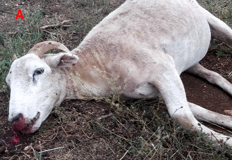
Figure 1. Gross pathology: (A) unilateral epistaxis
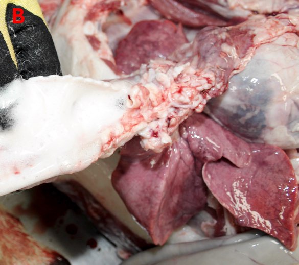
Figure 2. Gross pathology: (B) stable foam in the tracheal lumen
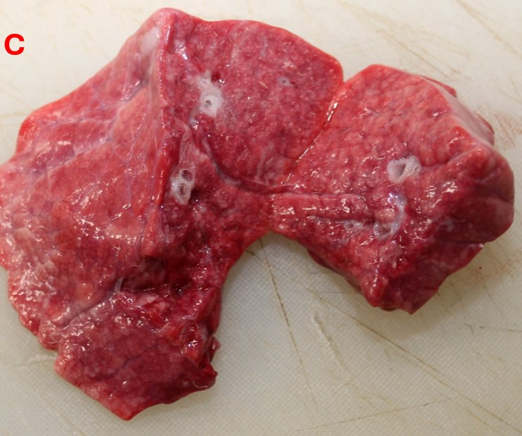
Figure 3. Gross pathology: (C) moderate diffuse interstitial pneumonia
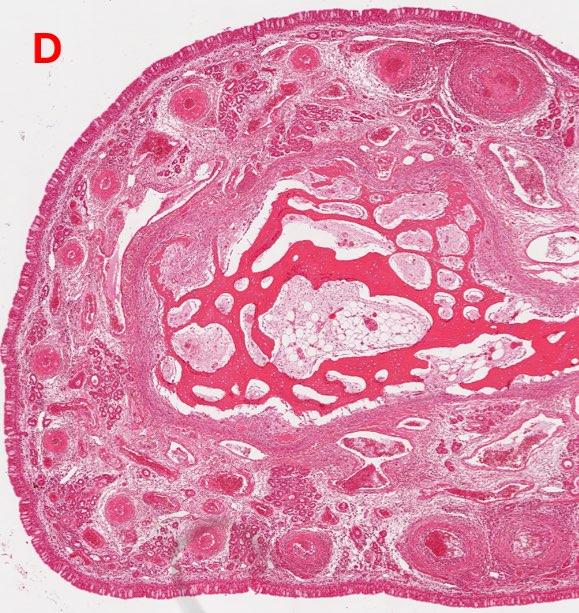
Figure 4. Histopathology: (D) multifocal, widespread vasculitis in the nasal turbinate
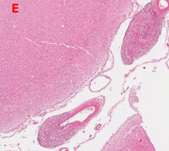
Figure 5. Histopathology: (E) multifocal vasculitis in the brain
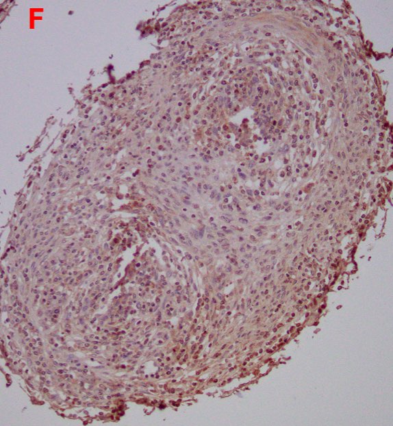
Figure 6. Immunohistochemistry: (F) affected vascular walls and infiltrating macrophages show strong immunohistochemical reactivity with anti-OvHV-2 antibody
DISCUSSION
Although OvHV-2 is traditionally regarded as a subclinical infection in sheep, there is increasing evidence that the virus can occasionally be associated with clinical disease and mortality in its natural host. Reported cases have described sheep presenting with weight loss, corneal opacity, epistaxis, rhinitis, neurologic signs, and sudden death, with histologic lesions characterised by multisystemic lymphoplasmacytic or lymphohistiocytic vasculitis resembling SA-MCF observed in susceptible species (Phillips et al., 2018; Sheehan et al., 2024).
Molecular investigations, including PCR and in-situ hybridisation, have demonstrated localisation of OvHV-2 nucleic acid within vascular lesions and infiltrating mononuclear cells, supporting an association between viral presence and lesion distribution (Pesavento et al., 2019; Saura-Martinez et al., 2021). Similar OvHV-2-associated systemic vasculitis has also been investigated in sheep in Ireland and Canada, further suggesting that clinical OvHV-2 infection in sheep may be under recognised (Sheehan et al., 2024; Ontario Animal Health Network, 2021).
These findings support recent reports that sheep are not uniformly asymptomatic reservoir hosts and indicate that OvHV-2-associated disease may express under certain conditions. Although MCF-like disease in sheep appears to be rare, the limited number of reports suggests it may be under recognised. This Australian case highlights the need for further investigation into the pathogenesis and epidemiology of OvHV-2 within the sheep industry.
Ethics Approval: The owner consented for use of data for teaching and research purposes (Veterinary Diagnostic Laboratory, Charles Sturt University).
REFERENCES
- Sheehan M, Pesavento PA, Campion F, Lynch J, McGettrick S, Toland B & Kennedy A (2024) First reported case in an Irish flock of MCF-like systemic necrotizing vasculitis in sheep associated with ovine herpesvirus 2 Irish Veterinary Journal 77(1): 7 doi.org
- Saura-Martinez H, Al-Saadi M, Stewart JP & Kipar A (2021) Sheep-associated malignant catarrhal fever: Role of latent virus and macrophages in vasculitis Veterinary Pathology 58(2): 332-345 doi.org
- Li H, Cunha CW & Taus NS (2011) Malignant catarrhal fever: Understanding molecular diagnostics in the context of epidemiology International Journal of Molecular Sciences 12(10): 6881-6893 doi.org
- Pesavento PA, Cunha CW, Li H, Jackson K & O'Toole D (2019) In situ hybridization for localization of ovine herpesvirus 2, the agent of sheep-associated malignant catarrhal fever, in formalin-fixed tissues Veterinary Pathology 56(1): 78-86 doi.org
- Makoni GM, Gerspach C, Fischer N, Rosato G, Fabian R, Grest P & Kipar, A (2024) Malignant catarrhal fever in a goat: Manifestation of virus-induced erythema multiforme Journal of Veterinary Diagnostic Investigation 36(2): 243-247 doi.org
- Phillips IL, Cunha CW, Galbraith D, Highland MA, Bildfell RJ & Li H (2018) High copy number of ovine gammaherpesvirus 2 DNA associated with malignant catarrhal fever-like syndrome in a lamb Journal of Veterinary Diagnostic Investigation 30(4): 623-627 doi.org
- Ontario Animal Health Network (2021) Investigation of ovine herpesvirus-2 as the cause of an idiopathic fatal vasculitis syndrome in Ontario sheep. University of Guelph