CASE NOTES
Nervous listeriosis in goats: Description of a clinical case in northern Spain
Itziar Montuega1, Miriam Martín1, Héctor Puyal1, Elena Longarón1, Karla Kostadinova1, Roberto Vitaller1, Janire Fernández2, David Guallar1,3, Pablo Quílez1,3, Delia Lacasta1,3, María Climent1,4
- Servicio Clínico de Rumiantes, Facultad de Veterinaria, Universidad de Zaragoza
- Belardi Albaitari Zerbitzuak S.L., Hernani, España
- Departamento de Patología Animal, Facultad de Veterinaria, Universidad de Zaragoza
- Departamento de Anatomía, Embriología y Genética Animal, Facultad de Veterinaria, Universidad de Zaragoza
Posted Flock and Herd 2026
Introduction
Listeriosis is an infectious disease of bacterial origin caused by Listeria monocytogenes, a Gram-positive, facultative intracellular microorganism that is widely distributed in the environment. This bacterium can survive under adverse conditions such as low temperatures, acidic pH, and high salt concentrations (Farber & Peterkin, 1991; Low & Donachie, 1997). Furthermore, this pathogen is of considerable importance in animal health and public health due to its zoonotic nature and its association with contaminated food.
In small ruminants, disease is characterised by low morbidity and high mortality, occurring sporadically or in outbreaks associated with predisposing factors (Center for Food Security and Public Health, 2007). The primary route of infection is ingestion of contaminated material, with poor-quality silage, particularly that with a pH above 5.5, being the most common source of infection (Radostits et al., 2007; Constable et al., 2021). Following ingestion, the bacterium invades the oral epithelium and may ascend via the trigeminal nerve to the brainstem, where it produces characteristic lesions of rhombencephalitis (Low & Donachie, 1997).
Clinically, listeriosis in small ruminants may present in several forms, the most common being the nervous or encephalic form, characterised by depression, head tilt, unilateral facial paralysis, circling behaviour, and progression to recumbency and death (Constable et al., 2021; Merck Veterinary Manual, 2023). Other less common forms include the septicaemic form, mainly in young animals, and the reproductive form, associated with abortions in the last third of gestation (Macleod et al., 1974). It is also worth noting that abortions caused by Listeria ivanovii have been observed in Australia.
The disease shows a higher incidence during colder periods, coinciding with the use of silage, and typically affects a small number of animals within a flock, albeit with a high mortality rate (Radostits et al., 2007). L. monocytogenes is transmitted to humans through the consumption of contaminated food and is therefore a significant food safety issue (World Health Organization, 2018).
Clinical case
The case occurred on a dairy goat farm located in the province of Guipúzcoa, in northern Spain, with an approximate herd size of 140 French Alpine goats. The animals were pregnant and managed under a semi-intensive system. Their diet consisted of hay, concentrate with mineral supplements, and silage, the latter having a high moisture content.
According to the farmer, the first cases appeared sporadically approximately one week prior to consultation, with animals showing acute-onset neurological signs and rapid progression to death within 48-72 hours. In total, four goats were affected consecutively (Figure 1), following a 'drip mortality' pattern. The farmer reported that the affected animals were those in the best body condition within the herd. Three of them died on the farm.
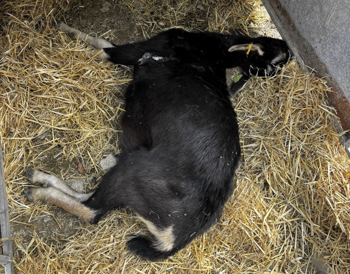
Figure 1. Goat recumbent on the farm following acute-onset neurological signs
The fourth case developed neurological signs the day prior to referral, presenting with recumbency and inability to stand. The animal was admitted to the Ruminant Clinical Service (SCRUM) at the Faculty of Veterinary Medicine of Zaragoza (Spain) the day after the onset of clinical signs.
The goat showed a depressed mental state, recumbency with inability to stand, marked sialorrhoea, presence of ulcers on the oral and vaginal mucosa, and pronounced ruminal hypomotility. Mild respiratory signs were also observed, consisting of bilateral serous nasal discharge and a productive cough, likely secondary to recumbency. Physiological parameters, including body temperature, heart rate, and respiratory rate, as well as mucous membrane status and body condition, were within normal ranges.
A blood sample was collected for a leukogram. The white cell profile showed a pattern consistent with acute inflammation, characterised by marked neutrophilia together with lymphopaenia, while the total leukocyte count was within the upper limit of normal, consistent with systemic inflammation of infectious origin.
Given the presence of signs suggestive of neurological involvement, a detailed neurological examination was performed. The animal was mentally depressed and unable to stand, preventing assessment of gait. Regarding spinal reflexes, tail tone and voluntary movement were preserved, whereas the anal sphincter and vulvar reflexes were absent in response to nociceptive stimulation. The myotatic reflex could not be adequately assessed; however, quadriceps muscle mass and tone were appropriate, suggesting functional integrity of the femoral nerve and spinal cord segments L4-L6. Withdrawal reflexes were present in both hindlimbs, requiring greater stimulus intensity on the left side, and no crossed extensor reflex was observed. The cutaneous trunci reflex was bilaterally intact.
In the cranial nerve examination, the olfactory nerve was not assessed. The pupillary light reflex was present bilaterally, and the menace response was intact, indicating functional visual pathways and cranial nerves II, III, and VII. Response to light stimulation elicited movement of the third eyelid, consistent with cranial nerve VI function. Regarding the trigeminal nerve (cranial nerve V), the mandibular branch was functional, whereas the maxillary and ophthalmic branches showed reduced response on the left side. The facial nerve (cranial nerve VII) exhibited partial left-sided paralysis with facial asymmetry and sialorrhoea (figure 2). Evaluation of the vestibulocochlear nerve (cranial nerve VIII) revealed a left ventrolateral strabismus upon head position change, consistent with a left-sided vestibular syndrome. The glossopharyngeal, vagus, accessory, and hypoglossal nerves (cranial nerves IX-XII) were functional.
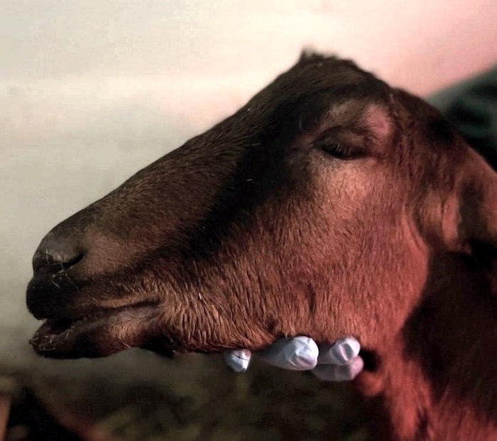
Figure 2. Goat referred to SCRUM showing partial left facial nerve (cranial nerve VII) paralysis and sialorrhoea
The differential diagnosis list was established based on the neurological findings, prioritising central processes with lateralised presentation. Small ruminant lentivirosis in its nervous form was considered but ruled out due to its chronic and progressive course. Polioencephalomalacia due to thiamine deficiency was also included, although it is typically associated with characteristic cortical lesions and a less focal pattern in the brainstem. Toxic plant intoxications were considered unlikely due to the limited number of affected animals and their typically bilateral and symmetrical presentation, while trauma did not explain the clustered occurrence of cases. Regarding the respiratory and digestive signs, conditions such as the caprine respiratory complex, aspiration pneumonia, and ulcerative stomatitis were considered, being interpreted as secondary or concurrent processes.
Integrating the clinical findings, the condition was concluded to be an acute, progressive, and asymmetrical process localised to the left brainstem, compatible with an inflammatory or infectious aetiology. The clinical presentation, disease course, and epidemiological context, particularly the association with silage consumption and the presence of head tilt and facial paralysis, strongly supported encephalic listeriosis as the most likely differential diagnosis.
Due to the unfavourable progression, the animal was euthanised and a necropsy was performed. Macroscopically, meningeal hyperaemia and cerebral oedema were observed. Histopathological examination revealed focal suppurative meningoencephalitis in the brainstem, with microabscesses, necrosis, and mononuclear perivascular cuffing (Figure 3), lesions highly suggestive of L. monocytogenes infection. Microbiological confirmation was achieved through isolation of the organism from brain tissue culture.
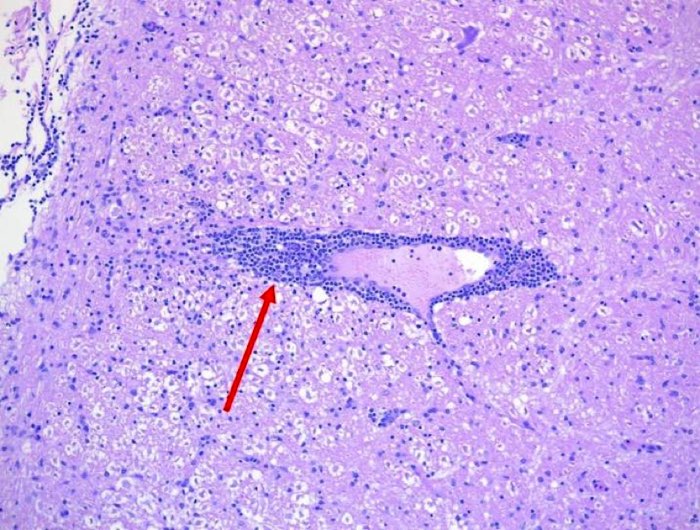
Figure 3. Histological section of the brainstem stained with haematoxylin and eosin (H&E), showing perivascular cuffing characterised by the concentric accumulation of mononuclear inflammatory cells around blood vessels (red arrow)
Discussion
Listeriosis in small ruminants is a disease of major importance, closely linked to the feeding of silage. The ingestion of poorly preserved or contaminated forage constitutes the main risk factor, promoting the proliferation of L. monocytogenes and its entry into the host. Silage pH is a critical factor: values below 4.5 limit bacterial multiplication, whereas values above 5.5, together with anaerobic conditions and suitable temperatures, favour its growth. Additionally, contact of feed with soil, inadequate bale storage, and contamination with faeces from birds or other animals act as additional sources of infection. The removal of superficial layers of silage from the silo and the discarding of mouldy areas reduces the risk, reinforcing that silage control is the fundamental preventive measure (Low & Donachie, 1997).
Although abortive and septicaemic forms also occur, where the bacterium invades the bloodstream and reaches the placenta, causing abortion, the cases described here correspond to the nervous form. The main route of entry for this form is oral, although nasal and conjunctival routes have also been described. After entering the host, the pathogen ascends to the brain via the trigeminal nerve (cranial nerve V), causing infection and inflammation, which explains the predominant localisation of lesions in the brainstem and the observed clinical signs (Low & Donachie, 1997; Oevermann et al., 2010). Perivascular cuffing represents one of the most relevant and characteristic histopathological findings, reflecting the inflammatory response of the central nervous system to bacterial infection.
The incubation period is typically two to three weeks but may extend up to two months. This long incubation period impacts case investigation and resolution: when clinical signs appear, animals may no longer be consuming the contaminated feed and new cases may continue to arise even after its withdrawal. Clinically, the neurological form predominates, with rapid progression, usually between one and five days from the onset of signs, and high mortality. Initial signs include depression, anorexia, reduced milk production, and transient fever progressing to incoordination, hemiparesis, unilateral facial paralysis, head deviation, and head tilt. In many cases, death results from secondary complications such as peracute pneumonia or enterotoxaemia.
Presumptive diagnosis requires a detailed history focused on feeding practices and the presence of compatible neurological signs. Definitive confirmation requires isolation and identification of the pathogen by microbiological culture or PCR. In the nervous form, the sample of choice is the brainstem from deceased animals, although spinal cord or cerebrospinal fluid may also be used, where histopathology can confirm the diagnosis. Serological tests have low sensitivity and specificity and are therefore not considered the diagnostic test of choice.
Treatment is challenging and often ineffective, particularly in advanced stages. The likelihood of success increases if therapy is initiated very early and with appropriate antimicrobial selection, taking into account the difficulty of crossing the blood-brain barrier. Intrathecal administration may improve drug distribution but is rarely feasible under field conditions. Some effective antibiotics include penicillin G or ampicillin at high doses (22,000 U/kg every 12 hours) or oxytetracycline, particularly in the early stages of the disease. Supportive therapy includes correction of dehydration and acidosis through intravenous fluid therapy and sodium bicarbonate. The administration of vitamin B1 (thiamine) is also recommended as it may contribute to clinical improvement in affected animals (Constable et al., 2021; Braun et al., 2002).
From a public health perspective, L. monocytogenes is a highly relevant zoonosis. Although morbidity in humans is relatively low, mortality in systemic or encephalitic forms may reach 20-30%, with hospitalisation rates exceeding 95% in Europe and the elderly, pregnant women, neonates, and immunocompromised individuals at most risk of severe disease (World Organisation for Animal Health, 2023). Transmission is mainly associated with ready-to-eat foods and contaminated products, including unpasteurised dairy products (Center for Food Security and Public Health, 2007). It is also a zoonotic risk to farmers and veterinarians delivering aborted foetuses.
Overall, listeriosis is a disease for which prevention, based on rigorous silage and feeding management, clearly outweighs treatment as a control strategy as listeriosis has both production and public health impacts.
Bibliography
- Abutarbush SM (2010) Book review: Veterinary medicine: A textbook of the diseases of cattle, horses, sheep, pigs and goats (10th ed.) The Canadian Veterinary Journal 51(5): 541
- Braun U, Stehle C, & Ehrensperger F (2002) Clinical findings and treatment of listeriosis in 67 sheep and goats The Veterinary Record 150(2): 38-42 doi.org
- Center for Food Security and Public Health (2007) Listeriosis. Iowa State University, College of Veterinary Medicine
- Constable PD, Hinchcliff KW, Done SH, & Grünberg W (Eds.) (2021) Veterinary medicine: A textbook of the diseases of cattle, horses, sheep, pigs and goats (11th ed.) Elsevier
- Farber JM, & Peterkin PI (1991) Listeria monocytogenes, a food-borne pathogen Microbiological Reviews 55(3): 476-511 doi.org
- Low JC, & Donachie W (1997) A review of Listeria monocytogenes and listeriosis The Veterinary Journal 153(1) 9-29 doi.org
- López-Almela I, Sheth CC, Gomis J, Gómez-Martín Á, Lecuit M & Quereda JJ (2025) Epidemiology, clinical and pathological features and outcomes of listeriosis in ruminants: a systematic review and meta-analysis The Veterinary Quarterly 45(1): 2598257 doi.org
- Merck Veterinary Manual (2023) Listeriosis in animals. Merck & Co., Inc. www.merckvetmanual.com
- Oevermann A, Di Palma S, Doherr MG, Abril C, Zurbriggen A & Vandevelde M (2010) Neuropathogenesis of naturally occurring encephalitis caused by Listeria monocytogenes in ruminants Brain Pathology 20(2): 378-390 doi.org
- Radostits OM, Gay CC, Hinchcliff KW & Constable PD (2007) Veterinary medicine: A textbook of the diseases of cattle, horses, sheep, pigs and goats (10th ed.) Saunders Elsevier
- World Health Organization (2018, February 20). Listeriosis www.who.int
- World Organisation for Animal Health (2023) Listeriosis www.woah.org