CASE NOTES
CALF DIPHTHERIA IN HEREFORD CALVES
Bruce Watt, Tablelands Livestock Health and Pest Authority, Bathurst
Posted Flock & Herd December 2011
Introduction
Fusobacterium necrophorum is a widespread anaerobic bacterium inhabiting the gut and excreted in the faeces of herbivores. The feet, oral cavity, pharynx, liver and occasionally mammary gland, abomasum, joints and female reproductive tract of ruminants can become infected, especially if damaged. Necrobacillosis typically affects the oral cavity (oral necrobacillosis) or larynx and pharynx (calf diphtheria) of calves raised under insanitary conditions or those subject to stress or concurrent disease (Beveridge 1983). Consequently, necrobacillosis is rare in beef calves raised on pasture.
Case report
Two calves approximately 2 months old in a mob of 25 Hereford cows with calves at foot were noticed to be sick. The dams, mixed age Hereford cows, were in fat condition and the calves were well grown. The mob was running on a barley grass, ryegrass and subterranean clover pasture with some improved species. The calves were initially examined on 20th April 2011. After calf diphtheria was diagnosed, the case was referred to a private practitioner for treatment. One calf appeared to improve with antibiotic (penicillin) therapy, but died suddenly on 15th May. The second calf recovered.
Clinical findings
At the initial examination, both calves were dyspnoeic. The first calf had an extended neck with open-mouthed breathing and stertorous respiration. The second calf was brighter but also exhibited snoring respiration. Respiratory sounds were most audible over the laryngeal. Inspiration could be obstructed by gently pressing on the lateral walls of the larynx. Both calves were febrile. Based on these findings, calf diphtheria was diagnosed.

Figure 1 - One affected calf died suddenly three weeks after antibiotic therapy was instigated, while the other recovered
Necropsy findings
The internal mucosa of the larynx was lined with a 5mm thick layer of yellow tightly adherent caseous material. Surrounding tissues were hyperaemic and some ingesta was lodged anterior to the diphtheritic ring within the larynx.
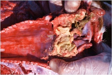
Figure 2 - Trachea and larynx opened to demonstrate diphtheritic membrane lining the larynx
Discussion
As the gross lesion was consistent with calf diphtheria it was considered unnecessary to confirm the presence of F. necrophorum.
This case is unusual in that it occurred in two otherwise apparently healthy beef calves raised under a conventional pasture system. It is suggested that the predisposing lesions are contact ulcers of the larynx caused by repeated closure of the larynx (Radostits et al. 2007). Previous respiratory disease may have contributed although there was no evidence of this in the herd.
Despite antibiotic therapy one calf died. Given the chronic and intractable nature of the lesion, this is not surprising. The calf would have been susceptible to asphyxiation either from necrotic material in the larynx or from ingesta.
References
- Beveridge WIB (1983) Animal Health in Australia, Volume 4 Bacterial Diseases of Cattle, Sheep and Goats, pp 178-183
- Radostits OM, Gay CC, Hinchcliff KW and Constable PD (2007) Veterinary Medicine, 10th Edition. p 1079