CASE NOTES
AN UNUSUAL PRESENTATION OF CONTAGIOUS ECTHYMA (SCABBY MOUTH/ORF) IN YOUNG RAMS
Libby Guest, District Veterinarian, North West LHPA
Posted Flock & Herd December 2012
Introduction
Seven of approximately 100, eight month old, homebred, Border Leicester stud rams were affected by an unusual presentation of contagious ecthyma (scabby mouth/orf) on a property near Narrabri, NSW.
The rams did not have the typically reported/cited lesions affecting the mucocutaneous junctions of the mouth and nose. Instead they presented with hyperkeratotic, proliferative papillary projections on the ears and dorsal cranium. One ram had a more typical contagious ecthyma lesion on its legs. The head and ear lesions were positive for Orf virus via electron microscopy.
History
Soon after entering an on- property feedlot for conditioning, 4 young rams from a mob of 100 were noticed to have moist, but "warty" skin lesions on their heads. A week later, 7 rams had lesions.
Clinical Signs
The four worst affected rams were presented for examination. All had markedly hyperkeratotic, wart-like projections, up to 15mm long and 2-5mm diameter affecting the dorsal cranium and ears (Images 1,2 & 3). The skin surface bled profusely when the projections were plucked. The owner reported that a week earlier, the lesions were moist and exudative.
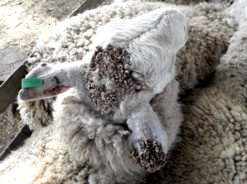
Image 1: Lesions on the head and ears

Image 2: Ear lesions

Image 3: Plucked hyperkeratotic skin lesions
None of the rams had lesions on the lips or nose. One of the rams had lesions on its lower front and hind legs that resembled contagious ecthyma (Image 4).

Image 4: Leg lesions more closely resembling contagious ecthyma. Unfortunately bleeding obscures much of the scabby lesion
Laboratory Findings
Lesions were taken from head/ear lesions on two rams and submitted to the NSW State Veterinary Laboratory Menangle.
Gram stain examination for Dermatophilus was negative on both samples.
Electron microscopy staining on samples both rams was positive for Orf virus.
Discussion
Radostits et al. describe contagious ecthyma lesions "as papules and then pustules, stages which are not usually initially observed, and progress to raised moderately proliferative area of granulation, and inflammation covered with a thick, tenacious scab". The lesions begin at the oral mucocutaneous junction and spread to the muzzle and nostrils. However the virus can enter the skin at any scarified areas and cause lesions. Image 5 depicts typical contagious ecthyma lesions from a different case (diagnosis also confirmed by electron microscopy).
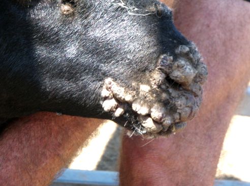
Image 5: Typical contagious ecthyma mouth lesions (Pic: Toni Jericho)
The case presented in this report is unusual in that the lesions were characterised by hyperkeratosis, and appeared on the dorsal cranium and ears, without lesions occurring near the mouth. There was no history of injury to the rams, grazing abrasive pastures (thistles) or ill-health in the mob of rams. Perhaps the virus entered via skin abrasions to the head following aggression between the rams in the feedlot. The owner reported that they had seen typical contagious ecthyma lesions in sheep on the property in the past, although the diagnosis was never confirmed. The owner indicated, during follow up discussions, that there were no new cases, nor any rams that developed more typical orf lesions around the mouth and nose. The affected rams recovered uneventfully within a couple of weeks.
Only one other case of orf virus causing hyperkeratotic lesions was found in the literature. Hooser et al. describe an atypical contagious ecthyma presentation in a sheep following a burn injury. In this case the lesions presented as an elevated, verrucous, irregularly surfaced area of tissue made up of numerous tightly packed 0.5mm diameter papillary projections. It is difficult to draw similarities between the two cases. The reason for the hyperkeratosis in either case remains a mystery.
References
- Radostits OM, Gay CC, Hinchcliff KW and Constable PD (2007) Veterinary Medicine, A textbook of the diseases of cattle, horses, sheep, pigs and goats. Saunders 10th Edition pp 1418-1421
- Hooser SB, Scherba G, Morin DE and Whiteley HE (1989) Atypical contagious ecthyma in a sheep after extensive cutaneous thermal injury Journal of the American Veterinary Medical Association 195(9)