CASE NOTES
Neurological Cases in Ruminants
Ted Irwin, District Veterinarian, North West LHPA, Warialda
Posted Flock & Herd September 2010
Case 1 - Swainsonine poisoning in goats
History and clinical signs
A private veterinarian phoned to report that a client had notified of neurological signs in her 3 goats. One goat was unable to rise and another was very wobbly on its feet. The owner did not think the third goat was affected but on inspection it had mild ataxia. The most affected two goats displayed a head tilt and nystagmus. The recumbent goat had some head tremor also. There was notable ataxia with a wide based stance in the most affected standing goat. When startled, the least affected goat toppled over.
The owner reported that the most recumbent goat was affected a year ago by the same symptoms but subsequently recovered until this year.
An inspection of the paddock revealed some shoots of Darling Pea (Swainsona sp.), access to various garden plants and shrubs, and a large area of rubbish and waste in which there were many sheets of lead protruding from a rusted old utility vehicle. The yard was only an acre in size and had been heavily grazed.
Available vegetation was sparse. No rock fern was seen but the area is known to contain rock fern which has thiaminase activity as well as the potential to cause a coagulopathy.
Differential diagnoses in this case were limited to lead poisoning, swainsonine toxicity and polioencephalomalacia (PEM). The owner noted that she had seen the goats feverishly going after the Darling Pea plants available.
post-mortem and pathology findings
Initially blood was sent to the lab for a lead assay. No other tests were carried out. Lead levels were normal. An autopsy was then carried out on the worst affected goat. Unfortunately the lab was unable to confirm swainsonine toxicity. There were no characteristic vacuolation of the neurones in the CNS. There was an unidentified pigment in the brain tissue and there was vacuolation of the hepatocytes. Some changes of minor note were reported in the kidney. No other tissues were examined.
Discussion
Swainsonine is a specific inhibitor of lysosomal -mannosidose causing accumulation of mannose in lysosomes (O.M. Radostits). This accumulation leads to vacuolation of the neurons of the CNS. Signs usually appear after 6-8 weeks of ingesting the plant.
Clinical signs include:
- Hind limb weakness with an inability to extend the hocks and an intermittent weaving gait
- Nystagmus
- Vestibular syndrome
- Wide-based stance
- Ataxia
- Disuse muscle atrophy
- Head-pressing, but not as severe as in PEM or lead poisoning
- Head tilt
- Head shaking
- Star gazing (in sheep)
- Proprioceptive deficits
- Startled goats may stumble or collapse
- Progressive paresis and ataxia to eventually involve the forelimbs
- Progressive weight loss
In goats they are usually bright and alert until a few days prior to death. Sheep almost always show profound depression.
Necropsy lesions are minimal. Emaciation and abomasal ulceration are the only gross lesions of note.

Figure 1. Sheets of lead visible

Figure 2. Only sparse feed available Dec 2009

Figure 3. Darling pea in full flower
Despite the inconclusive lab results, a presumptive diagnosis of swainsonine toxicity was made. A subsequent visit to the property has revealed that the disease has progressed somewhat in the two remaining goats although they are both still bright and eating well.
Abundant Darling Pea has grown after good rains in late December adding to the likelihood of the diagnosis.
One diagnosis not given much consideration in this case is Coonabarabran staggers from chronic grazing of Tribulus sp. (yellow vine, cat head, caltrop). This plant was present in the area in significant amounts and owing to the scarcity of better vegetation was probably grazed to a certain extent.
Case 2 - Lead poisoning
History and clinical signs
A producer phoned to report two yearling steers that had jumped a fence into a cultivation paddock and were now showing signs of depression and blindness. He was unsure how long they had been present in the paddock. The paddock was black soil with some growth of lucerne and regrowth of oats.
Both steers were able to be approached in the paddock and were apparently blind with absent menace responses. They had some drool from the mouth and mild nasal discharges.
Differential diagnoses included lead poisoning and PEM. Injections of thiamine were given intramuscularly and blood was taken from the tail vein. One steer was circling.
A repeat visit was conducted the next day as one of the steers was recumbent. There was no response to thiamine injections. The recumbent steer was euthanased and autopsied.
Background
A thorough post-mortem was not conducted. Large amounts of lead were found in the reticulum and therefore a diagnosis of lead poisoning was made. The second steer was then examined and was frothing at the mouth and nodding its head in a recurrent nervous 'tick' fashion. It was also euthanased by firearm. The source of lead was not identified but there was a large area of ash deposits from a wood fire which may have contained a source of leade.g.lead based paint.
Discussion
Clinical signs of lead poisoning include:
- Posterior paresis in lambs
- Absent menace responses
- Head pressing
- Blindness
- Fasciculations and tremor
- Convulsions
- Sudden death
Blindness has generally been the most reported sign, followed by sudden death. Rarely, convulsions and frothing at the mouth are seen.

Figure 4. Steer affected by lead

Figure 5. Steer affected by lead

Figure 6. Lead in the reticulum
Case 3 - Polioencephalomalacia
History and clinical signs
A producer phoned to report 2 heifers that were wandering aimlessly and appeared depressed and possibly blind. I visited the property and inspected the cattle that were slow to react to visual stimuli and had absent menace responses. One was head pressing against a fence. Complete blindness could not be confirmed as they were able to be moved into the yards and crush without too much trouble.
Both animals exhibited a fever of over 40C but there were no other obvious clinical signs.
Differential diagnoses included PEM, Sporadic Bovine Encephalomyelitis (SBE), lead poisoning, and meningitis.
On inspection of the paddock there was abundant turnip weed (Brassica sp.) growing and in flower. It had been heavily grazed by the cattle in this paddock.
Clinical pathology
Lead was ruled out by blood tests. Chlamydial titres were positive but no follow up titres were taken to confirm active infection. However, there was no joint swelling in either heifer to indicate SBE. The case was followed up a week later with a phone call and the heifers had improved marginally. The case was not followed up further than this and so an autopsy was never carried out to confirm PEM.
Discussion
The clinical signs of PEM include:
- Nystagmus
- Opisthotonus
- Head pressing and head twitching
- Menace responses are usually diminished or absent
- Blindness
Clinical signs can mirror those caused by lead poisoning and so it is difficult to differentiate the 2 disorders. In the Northern Slopes region of the North West LHPA it is very unusual to see PEM from bacterial changes to the rumen causing breakdown of rumenal thiamine. PEM in sheep can be associated with ingestion of rock/mulga fern (Cheilanthes sieberi), which also causes a coagulopathy. PEM in cattle is more commonly associated with ingestion of large amounts of wild turnip (Brassica sp.) or iatrogenic sulphur toxicity through ill-prepared dry lick supplements.
There is debate over whether thiamine injections will assist in cases of PEM caused by sulphur intoxication. Even in cases of thiaminase induced PEM there is variability in the response to thiamine injections and therefore the effect in sulphur induced cases would be difficult to measure. Sulphide inhibits cellular respiration leading to hypoxia which may be sufficient to create neuronal necrosis in PEM. The CNS lesions are indistinguishable from those in the naturally occurring disease.
Rumen bacteria take approximately 10-12 days to adjust to diets high in sulphur before becoming capable of producing toxic concentrations of sulphide in the form of hydrogen sulphide.
If animals are removed from the toxic source then the lesions are reversible but only in the early stages. Once the disease has advanced beyond a few days exhibiting clinical signs then the scope for recovery is minimal.
Case 4 - Pregnancy toxaemia (fatty liver syndrome) in cattle
History and clinical signs
In April 2009 a producer phoned to report a number of cattle deaths on his property north west of Walgett. All dead cattle were pregnant heifers in deteriorating condition and the heifer mob had recently been separated from the cow mob to a new paddock with less available feed.
The clinical signs described were:
- Sudden death
- Weakness or paralysis
- Muscle spasms
- Muscle tetany
- Loss of condition
- Frenzy/agitation
On examination of a mob of yarded heifers it was noted that some of the heifers were very stirred up and agitated at being moved into the race and crush. The owner noted they had lost a lot of condition recently and mentioned that many were wandering aimlessly around the fence line and not grazing. The symptoms also seemed to be exacerbated by mustering.
One heifer in particular was showing signs of muscle fasciculations and was quite agitated. She had a fever of 41C but there was no nystagmus and the menace response was decreased. Approximately 50mls of thiamine was given 'steadily' into the jugular vein. Immediately after this injection the heifer seemed to become weak and was let out of the crush at which time the animal collapsed on its front legs and lay on its brisket with back legs holding the rear end off the ground. The heifer exhibited signs similar to a spastic paralysis and then it toppled over and lay on the ground with all four legs stretched out laterally. After 30 seconds it made an effort to rise which was successful and then it staggered a few metres in a very stiff-legged gait. The animal subsequently collapsed again and lay in this position for about 2 minutes. After which time it rose without incident and walked off seemingly unhindered.
The owner described a similar episode of collapse while mustering the heifers at which time the collapsed heifer died suddenly.
The differential diagnoses for these signs included:
- Lead poisoning
- Pregnancy toxaemia
- PEM
- Flood plain staggers
- Billy-button poisoning
- Hypocalcaemia
- Hypomagnesaemia
- Other toxicity
- Tetanus
Clinical pathology
Hypocalcaemia and hypomagnesaemia seemed unlikely given the conditions and these were ruled out on blood tests. Lead poisoning was ruled out by blood tests and there were high levels of ketones in the blood indicating that pregnancy toxaemia was the most likely diagnosis. The heifer was moved to a better paddock but died 2 days later. A post-mortem revealed a massively inflamed liver, two times the normal size with marked yellow discolouration of the liver and of the whole carcass. Histopathology confirmed a massive fatty infiltration of the liver. The brain was not removed for histology.
Discussion
Firstly, the spectacular clinical signs observed here may well have been the result of an anaphylactic reaction to administration of i.v. thiamine. The fact that similar signs were reported in another dead heifer points more towards hepatic encephalopathy. Thiamine is recommended to be given intramuscularly and is a registered product for use in dogs and horses.
The long list of differentials including a couple of plant toxicoses makes this a difficult case to narrow down. The lack of diagnostic blood tests to rule in or out certain diseases also adds to the difficulty.
In an unwell animal that may be affected by PEM or other condition and is heavily pregnant may well show a higher than normal level of ketones as a result of starvation. The history of aimless wandering combined with the high temp suggested that PEM may well be the diagnosis. It would be ideal to have access to a quick blood test for the condition but this is not possible. Lead was also high on the list but is easy to rule out. In this case there were diminished menace responses just as with lead and PEM. The episode of collapse could easily fit with the convulsions seen in lead poisoning.
Billy buttons, or plains plover daisy (Ixiolaena brevicompta) has been known to cause death in sheep.
The signs seen with intoxication from consumption of the mature seed head are:
- Sudden death: animals that are disturbed run a short distance (10-30m) then suddenly collapse and die
- Tiring syndrome: animals are barely able to exercise and show hind leg weakness and staggering - some collapse and die, others recover. Recovery may take up to 14 days.
Signs of the disease appear first in lambs 3-6 months of age within 7 days of starting to graze pasture heavily infested with billy buttons. Adult stock show signs of the disease after a further 7-14 days. The disease is believed to be caused by crepenynic acid, an unusual fatty acid present in the seed of billy buttons.
There is no reference to the disease in cattle but they are also susceptible (W. Hetherington, personal communication 2009).
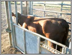
Figure 7. Heifer with pregnancy toxaemia

Figure 8. Marked fatty infiltration of the liver
General Discussion
Although all four cases are of distinctly different aetiologies, they presented with a number of similar clinical signs as well as other unique signs. The similarities are obviously indicative of the fact that all the conditions have affected the CNS to some degree and thus eliciting pathologies of the upper motor neurone system. To distinguish them in the field can be quite tricky.
Animals affected by lead poisoning can present with signs of frenzy or convulsions, they often present as quite depressed and with a reduced reaction to stimuli, almost identical to cases of PEM.
Similarly, advanced cases of lead and PEM may well present with nystagmus in the latter stages but in the opinion of the author this is rarely the case and is likely to be an excellent way to distinguish these disorders from those more commonly associated with nystagmus such as swainsonine toxicity and Coonabarabran staggers. These latter diseases more commonly present with vestibular syndromes that cause ataxia, a wide based stance, and head or ear twitching.
Pregnancy toxaemia and fatty liver syndrome may rarely lead to extreme signs of muscle spasms and collapse but in this case the signs were consistent with numerous other causes of neurological disease. The key to a cost efficient diagnosis here was purely based on history and signalment which highlights the importance of using the history as well as carrying out an in depth clinical examination and property inspection.
References
- Barbosa RC, Riet-Correa F, Lima EF, Medeiros RMT, Guedes KMR, Gardner DR, Molyneux RJ, De Melo LEH. Experimental swainsonine poisoning in goats ingesting Ipomoea sericophylla and Ipomoea riedelii (Convolvulaceae). Pesq. Vet. Bras. 2007; 27(10):409-414
- McKenzie R. Toxicology for Australian Veterinarians. CD 2002
- Radostits O, Gay C, Blood D, Hinchcliff K. Veterinary Medicine, 9th Ed. W. B. Saunders, 2000
- Smith MC, Sherman DM. Goat Medicine, 2nd Ed. Wiley-Blackwell 2009
- en.wikipedia.org