CASE NOTES
BLACKLEG IN CATTLE: A RETROSPECTIVE
Keith Hart, SDV Cumberland
Posted Flock & Herd March 2011
Introduction
Blackleg is a gangrenous myositis caused by the bacterium Clostridium chauvoei, which is commonly found in the soil and in the gut contents of normal cattle. It has a spore phase which is highly resistant to environmental conditions in soil and is known to persist for many years. In cattle, the disease occurs mainly in young animals 6-24 months of age in good nutritional condition – it is commonly observed that the best animals succumb.
An interesting feature of blackleg is that, in spite of the disease being officially recorded in NSW for over 130 years:
The pathogenesis of the disease is uncertain (2)
The same authors, two of the professions leading veterinary pathologists, wrote 37 years later:
The detailed pathogenesis of blackleg is still somewhat uncertain (7)
The best description of blackleg pathogenesis in cattle that I have seen is also the most recent, in Diseases of Cattle in Australasia (8). The authors state (p788) that:
The sequence of events culminating in a clinical blackleg incident is still not completely known. It is assumed that Cl. chauvoei spores are ingested and cross the intestinal wall. As a result spores become freely distributed to a variety of tissues including muscle. The spores are harmless to the animal until wounds, trauma or bruising damage the muscle tissue in such a way that an environment is created that is favourable for the spores to begin their vegetative growth.
In this paper I will present:
1. A data set of 45 laboratory confirmed cases of blackleg in cattle between 1973 & 2002 (confirmation was by histopathology and/or fluorescent antibody test for Clostridium chauvoei). The cases all occurred within the old Moss Vale Rural Lands Protection district, comprising the regions of Sydney Basin, Illawarra Coast (Wollongong to Nowra) and the Southern Highlands.
2. A case of major outbreak of blackleg in a Sydney Basin feedlot, where anthrax was the differential diagnosis.
3. A case of a ‘classic outbreak’ of blackleg on a Southern Highlands property where the owner never vaccinated, but the disease had never been seen in living memory.
Analysis of the Camden office blackleg files
Hard copy files at Camden documenting laboratory confirmed blackleg cases in cattle go back to 1973. Veterinarians from the Rural Veterinary Centre at the University of Sydney, Cobbitty, investigated the majority of these. The RVC had an active large animal practice in the Camden district at least between 1973 and 1986.
Seasonal incidence of blackleg.
The majority of cases occur in late spring/early summer and in autumn (10). This seasonal incidence is commonly referred to in the Australian literature.
Camden figures:
| Season | Number of outbreaks |
|---|---|
| Dec – Feb | 21 (45%) |
| Mar – May | 4 (9%) |
| Jun – Aug | 11 (23%) |
| Sep – Nov | 11 (23%) |
In this district, autumn is clearly the lowest risk period, in contrast to the observations in the literature. Essentially, at least in this district, blackleg in cattle can occur at any time of year – every month had at least one outbreak recorded against it.
Age/vaccination status
Many records did not indicate the vaccination status or the ages of the cattle that died. The following figures show cases where age, vaccination status and number of deaths were recorded. There is necessarily some overlap in the age ranges.
| Age range (months) | Number of deaths | Number of outbreaks |
|---|---|---|
| 3-4 | 7 | 2 |
| 3-8 | 3 | 1 |
| 4-5 | 15 | 5 |
| 4-9 | 3 | 1 |
| 5-14 | 3 | 1 |
| 6-8 | 20 | 8 |
| 8-10 | 6 | 4 |
| 11-24 | 2 | 2 |
All of the deaths recorded above were in unvaccinated cattle.
In only two outbreaks did the history record any vaccination – three animals between 12 and 24 months of age died of blackleg after receiving a single vaccination as a calf. This is consistent with field observations and the literature.
Effect of rainfall.
In some areas there is an increased prevalence in areas of high rainfall. (5).
An extraordinary proportion of blackleg outbreaks on file were investigated by the RVC, Cobbitty in the summer of 1974/1975. Twelve of 45 (27%) cases were investigated during this short period. To put it another way, there were 120 seasons in the 30 years of data studied – 1 season (0.8%) accounted for 27% of the blackleg cases documented.
1974 was also one of the wettest years in living memory in Eastern Australia. It is reasonable to speculate that this spike in blackleg cases was associated with exceptionally wet conditions experienced during 1974 (the 1974 rainfall total for Sydney has not been exceeded since then).
Distribution of lesions.
Many of the cases enthusiastically investigated by the RVC veterinarians in 1974/1975 indicated the sites of the lesions (they probably had students with them and spent more time searching than a practitioner or DV would normally do). Twelve cases had the sites of blackleg lesions described, and six of 12 had lesions in >one site (the largest number was four sites). The following were described as typical blackleg lesion sites:
| Site of lesions | Number |
|---|---|
| Brisket | 1 |
| Neck | 1 |
| Front limb/shoulder | 4 |
| Hind limb/hip | 8 |
| Tongue | 1 |
| Masseter | 4 |
| Psoas/long. dorsi | 1 |
| Abdomen | 1 |
| Myocardium | 1 |
Improving management
I took up the position as DV Camden in May 1987, and have always pushed routine clostridial vaccination of calves (two doses), with a booster at 12 months of age. The following figures to some extent reflect the increasing district use of clostridial vaccines in cattle, but are also influenced by such factors as the increasing cost of calling veterinary practitioners to investigate cattle mortalities, the fluctuating value of cattle and the increasing proportion of hobby farmers. A number of factors therefore influences the reduction in number of outbreaks, but the increased use of clostridial vaccines is certainly one of them.
| Period | Number of outbreaks investigated | Number / year |
|---|---|---|
| 1973-1975 (3 yrs) | 20 | 6.7 |
| 1976-1986 (11 yrs) | 12 | 1.1 |
| 1987-2010 (23 yrs) | 15 | 0.7 |
The figures from the last 23 years are also influenced by the fact that I am comfortable in diagnosing blackleg in cattle based on history and autopsy findings, so there are few recent copies of laboratory submissions to contribute to the statistics. The actual number of blackleg outbreaks investigated between 1987 and 2010 would be at least twice that recorded.
Colleagues from the anthrax areas of NSW may find this approach challenging, but in spite of the initial diagnosis of anthrax being made in the Cumberland district in 1847, ‘Cumberland Disease’ has not been recorded here since before 1951 (1).
‘Post-mortem examination, provided it is carried out soon after death, provides confirmation when the characteristic lesions are found, although sometimes they may be detected only after an extensive search. Supporting evidence may be available from circumstances such as susceptible age, absence of a wound (wound entry is often associated with malignant oedema, but not blackleg), good body condition (I cannot recall ever diagnosing blackleg in an animal in store condition), season (but see comments above) and the fact that the animals are on pasture.’ (8)
CASE 1: MAJOR OUTBREAK OF BLACKLEG IN AN OPPORTUNITY FEEDLOT AT BADGERYS CREEK, SYDNEY BASIN
This large (for the Sydney Basin) opportunity feedlot contained 560 twelve to eighteen month old, mixed sex cattle being fattened for the export market. They were run on grass on a leased, future development property and supplemented in paddock feeders with a mixed ration including waste bread and pollard. Cattle are purchased as forward stores from the Camden saleyards. Routine clostridial vaccination was not practised.
Time line:
Sun 8/7/2007 - All cattle normal
Mon 9/7/2007 - Several cattle found dead. Relatively inexperienced peri-urban veterinary practitioner called in and diagnosed anthrax. DV away on leave. Managing Ranger (Andrew Glover) instructed by RVO to collect smears from all available carcasses for anthrax exclusion. Smears collected from 16 carcasses (more had died during the day). All smears negative for anthrax.
Tues 19/7/2007 – DV investigates outbreak and diagnoses blackleg. Five in 1 vaccination program initiated for all introduced animals. All cattle on farm vaccinated twice with 5 in 1. Mortalities stop prior to the second vaccine dose.
Autopsy
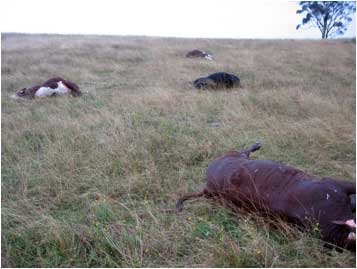
There were plenty of carcasses to choose from – 20 animals had died by this time, and five were sick and sitting down but still able to rise and walk away when approached. These animals were lame. Most carcasses were in the typical position – lateral recumbency with legs sticking up in the air, plus usually bloody discharge from one or more body orifices (usually the nose). However, two unusual cases were found dead in sternal recumbency with their legs straight out behind.
Most of the carcasses were over 24 hours old – over the years I have found autopsy of such cases unrewarding when blackleg is suspected, due to the overgrowth of post-mortem invaders – especially the summer months. However, one carcass was found that had died overnight and had been well preserved by the cold conditions although it was over 12 hours old. Typical subcutaneous gas accumulations and blackleg lesions in the muscle were found in the shoulder area.
Histopathology was consistent with clostridial cellulitis and myositis and was therefore malignant oedema and/or blackleg. Fluorescent antibody test on muscle impression smears was positive for Cl. septicum (malignant oedema) but negative for Cl. chauvoei (blackleg). However, this is entirely expected in a 15 hour old carcass – Cl. chauvoei are fastidious, and are rapidly outgrown by aggressive post-mortem invaders, including Cl. septicum.
Interesting points:
34 of 560 cattle eventually died from blackleg on this property (6% mortality). Losses were spread over no more than 10 days, and stopped before the second dose of 5 in 1 vaccine was given. The best-conditioned animals were invariably involved. This is the largest single cattle mortality due to blackleg that I have ever seen.
The case represented an interesting transition from regulatory to advisory – on Monday the case was suspect anthrax and the cattle were detained. On Tuesday, the anthrax results were negative and the diagnosis was blackleg, allowing the detention to be lifted and an advisory approach to be adopted.
The mortalities occurred during wet conditions (see above) and the property owners had been doing some earthworks in the paddock where most of the cattle died. An association between blackleg outbreaks and the excavation of soil is often mentioned in the literature.
All the sick cattle were given large doses of penicillin. Nineteen animals (>50% of the treated cattle) survived. While there is no way to be sure (given that all cattle were purchased from the Camden saleyards) I suspect that these animals had been vaccinated as calves and had some residual immunity, which with the antibiotics allowed them to recover. In my experience, blackleg in cattle with no immunity is invariably fatal.
At the end of the 2007 / 2008 financial year, the owner requested a letter from me documenting the cause of the loss of cattle during the year. The owner's accountant was concerned that writing off such an unusually large number would raise the suspicions of the ATO.
CASE 2: TYPICAL OUTBREAK OF BLACKLEG IN THE SOUTHERN HIGHLANDS
A loss of five out of seven, six to nine month old unvaccinated heifers was investigated in September 2010. Two calves were available for autopsy – one had died overnight (but there had been a frost), and the other had been dead for a maximum of 4 hours. The case provided a rare opportunity to obtain some fresh samples from a blackleg case.
The autopsy showed classic gross and histopathological lesions of blackleg, with typical oedema, emphysema and myositis. The comment from the RVL pathologist was:
Thank you for this great case. The histological findings are classic for blackleg, complete with intralesional bacilli.
Interesting elements of the history in this case were:
Early spring – see comments on seasonal incidence of blackleg above.
The owner (80 years old) had been on this property for 60 years, and had never seen anything like this before, and never vaccinated his cattle for clostridial disease.
While there were other mobs of susceptible age calves in other paddocks, the losses were confined to one paddock only. In this paddock, some timber had been piled up and the soil disturbed. Calves in particular and cattle in general are attracted to disturbed earth, and if blackleg spores were disturbed in this way, the blackleg outbreak has a possible explanation. Once again, the history of soil disturbance referred to in the literature crops up. Since this is a closed herd, the question is where did the spores come from.
Blackleg outbreak
While aspects of blackleg in cattle remain a mystery, the above cases illustrate that there is at least some consistency to the mystery of a disease that can appear and then disappear like a thief in the night.
References
- Albiston, H (1965) Seddon's Diseases of Domestic Animals in Australia – Bacterial Diseases (Part 5, Vol. 1) Commonwealth Department of Health
- Jubb, K & Kennedy, P (1970) Pathology of Domestic Animals, 2nd ed. Academic Press, New York & London
- Beveridge, W (1983) Animal Health in Australia – Bacterial Diseases of Cattle, Sheep and Goats (Vol. 4) Australian Bureau of Animal Health
- Hungerford, T (1990) Diseases of Livestock, 9th ed. McGraw Hill, Sydney
- Radostits, O et al. (2000) Veterinary Medicine 9th ed. W.B. Saunders, Sydney
- Hirsch, D et al. (2004) Veterinary Microbiology 2nd ed. Blackwell Publishing, Iowa, USA
- Jubb, K & Kennedy, P (2007) Pathology of Domestic Animals 5th ed. Academic Press
- Parkinson, T et al. (2010) Diseases of cattle in Australasia New Zealand Veterinary Association Foundation