CASE NOTES
Vitamin A deficiency in sheep and cattle in North Western NSW
Megan Davies, District Veterinarian, Narrabri, Shaun Slattery, District Veterinarian, Narrabri and Judy Ellem, District Veterinarian, Gunnedah
Posted Flock & Herd December 2019
Introduction
During drought conditions between 2014 and 2019, District Veterinarians investigated multiple cases of Vitamin A deficiency, presenting a wide range of clinical signs. Reported clinical signs of Vitamin A deficiency include anorexia, ill thrift, diarrhoea, blindness, convulsions, paralysis, recumbency, abortion or stillbirths, poor skin health, and immunosuppression manifesting as increased susceptibility to infectious diseases such as pneumonia, pinkeye and mastitis1,2,3. Many of these signs were observed in the field by District Veterinarians in the North West. Four of these cases will be discussed in detail in this paper, with reference made to the findings in other cases. These cases highlight the importance of monitoring Vitamin A levels in stock which present with an array of clinical signs. It also demonstrates the importance of educating producers regarding the importance of supplementation during prolonged periods of dry.
OVERVIEW
Vitamin A is required by sheep and cattle for a variety of functions throughout multiple body systems. These include maintenance of night vision, growth and development, immune function, fertility, and maintenance of epithelial tissue3,2,1,4,5. Congenital defects are common in the offspring of deficient dams, with affected calves showing blindness, nystagmus, weakness and incoordination2. The liver is the primary storage organ of Vitamin A2,6. Vitamin A levels in the blood are not representative of levels stored in the liver until these stores are so depleted that there is a concurrent fall in blood Vitamin A levels6.
Cattle can survive on stored Vitamin A for approximately six months before a deficiency results in clinical signs, while adult sheep may be on a deficient diet for 18 months before their stores are depleted and the disease becomes evident3. Younger sheep generally become deficient after 5-8 months, but may not show clinical signs for up to a year3. Calves and lambs are born with low Vitamin A levels, and rely on colostrum until Vitamin A is provided by the diet2.
Plants are poor sources of Vitamin A itself, but contain carotenoid precursors, which are converted to Vitamin A in the small intestine by enterocytes5. From there it is transported to the liver via the blood stream6,7,5. Deficiencies are generally primary deficiencies, arising from inadequate access to green feed, lack of adequate supplementation, or the breakdown of Vitamin A additives in commercial feeds3. Deficiency is common in cattle and sheep during drought conditions, or in stock being full hand fed in conditions such as feedlots3.
Over the period of July 2014 to December 2018, 37 properties in the North West had stock tested for Vitamin A and submitted to the NSW DPI State Veterinary Diagnostic Laboratory. Ten of 16 cattle herds tested had levels below laboratory reference ranges with three at marginal levels. Of 21 sheep flocks tested, eight had levels below reference ranges.
The range of clinical signs observed included weakness, recumbency, weight loss, ill thrift, eye infections, sudden deaths, poor coat condition, stillbirths, microopthalmia, diarrhoea, seizures, pneumonia and mastitis. In most cases, there were other contributing or primary conditions, including deficiencies in calcium, magnesium and energy.
Conditions across the North West of NSW between 2014 and early 2019 (the time of writing) were substantially drier than average. Though the region was not classified as "drought affected" until March 20188, many livestock enterprises had experienced limited pasture feed since late 2016. Many enterprises, especially in the more western areas had been providing increasing levels of hand feeding since early 2017. Figure 1 shows the intensity of the drought conditions in the North West of NSW in December 2018.

Figure 1. Drought conditions in North West NSW in December 20188
The most common feedstuffs used throughout the district were cereal grains (wheat and barley), white cottonseed, faba beans, and a variety of hay, straw and other low quality roughage (often sourced from other regions). Pellets were also commonly used for calves, lambs and early weaned stock.
Hay and silage can provide a source of Vitamin A in the absence of green pasture, however, both Vitamin A and its precursor carotenoids are easily destroyed by oxygen, heat, light and acids5. Significant losses occur during the process of preserving and storing fodder2. Any late-cut hay, bleached by sun or stored for long periods loses much of its carotene content3. Cereal grain is a poor source of Vitamin A3,6,5. Pelleting of feed can cause losses of up to 32% of the Vitamin A in the original feedstuff3. Losses of Vitamin A are further caused by heat, light and mineral mixes. The major source of Vitamin A used in supplements is trans retinyl acetate, which is made into beadlet form, with additives such as antioxidants9 to provide physical and chemical protection against adverse factors normally present in feed5. It is commonly used in mash and pelleted feed, however is still best used fresh, as studies have shown that Vitamin A levels diminish significantly when feed is stored - over 50% loss over one year in some instances5.
Case reports
Case 1 - Scruffy cows at Boggabri - September 2018
History
A mob of 130 Charolais X Angus cows with calves at foot were investigated for what the owner reported as "scruffy coats". Approximately 15 cows were affected. They were generally in poor body condition, averaging 1.5-2/5 body condition score (figure 2). They exhibited clinical signs of alopecia, which was predominantly around the eyes, on the neck (figure 3), backline and between the hind legs (figure 4). The lesions were not pruritic, and were characterised by scaly skin and loose hair which was easily pulled out in tufts (figure 5). The cattle were being fed white cottonseed and rice straw, with scant dry native pasture.
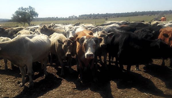
Figure 2 The affected mob, showing generalised poor body condition

Figure 3. Alopecia on the neck of affected cow

Figure 4. Widespread alopecia between the hind legs of an affected cow

Figure 5. Scaly skin and tufts of loose hair on the dorsal midline of an affected cow
Examination of the skin for lice, ticks, and any evidence of contact with an irritant such as a chemical was unrewarding. Skin scrapings were negative for mites.
Laboratory findings
Examination of skin scrapings at the NSW DPI State Veterinary Diagnostic Laboratory for Dermatophilus was negative, and fungal culture was similarly unrewarding.
Blood collected from three affected cows showed no significant abnormalities in biochemistry. Vitamin A test results showed low levels in two of the three cows tested (0.36, 0.18 and 0.20mg/L, normal range 0.26-0.6mg/L). There were no clinical signs or abnormalities in the calves present in the mob, suggesting that the deficiency had occurred recently, rather than throughout gestation.
Further cases
A second case on a property approximately 30km away was attended to in the same week by another District Veterinarian. The clinical signs were similar - alopecia with no pruritus. Tufts of hair were easily pulled from the epidermis leaving an area with scaly skin. Diagnostic testing in this case, including skin biopsies was similarly unrewarding. Vitamin A levels were not tested in this case, but in hindsight it is thought to be a likely diagnosis.
The location of the lesions in both cases was consistent with reported sites in other cases, being the head, neck and tail base7. Similarly, in other reported cases, no other abnormalities were noted, either clinically or on post-mortem examination, which was the case with the cattle observed in these cases.

Figure 6. Alopecia on neck of affected cattle at second property.

Figure 7. Marked Alopecia on the face of an affected cow on the second property
In the following two weeks another three similar cases were reported, within a 100km radius of the first case. In two of these cases, the owners had recently moved cattle onto green oat crops, and both reported an improvement in clinical signs since the move to green feed. It is notable that relatively nearby herds had all reached the level of deficiency required to show clinical signed at similar times.
Case 2 - Deaths in poddy lambs at Pilliga - July 2018
History
In July 2018 the Narrabri Local Land Services (LLS) office was contacted regarding ongoing deaths in poddy lambs on a Burren Junction property. Eventually 26 of 40 lambs died from an inappetence and ill-thrift syndrome due to Vitamin A deficiency. This investigation suggests the value of poddy lamb mobs in determining potential for Vitamin A deficiencies in paddock sheep and the response to provision of green feed as a quick diagnostic aid.
The poddy lambs were sourced from two mixed farming enterprises 30 km apart owned and managed by the same farmer. By July 2018 both properties had been without green feed for an extended period of time. On the first property this period was 18 months. On the second property a sporadic storm event generated green pick that reduced the period to 7 months. Lambs from both holdings had died. No other Vitamin A had been provided.
The sheep on both properties had been hand fed faba and cereal grain ration continuously for 12 months.
The poddy lamb mob was under the care of an experienced person who was able to attend the mob several times a day. The lambs were held in a well set out yard pen. They were fed 2 litres of lamb calf replacer per lamb each day, ad lib. commercial pellets, ad lib. oaten hay and some faba beans. Despite this, visually assessed growth rates were disappointing. Both the milk replacer and pellets noted the inclusion of Vitamin A on the packaging.
Clinical signs
At the initial visit the lambs were 3-9 weeks old. At this stage 16 lambs had died in the previous three weeks. Deaths were described as sudden and quiet, often with the lamb being observed normal at feeding and then found dead a few hours later.
Clinically the poddy lambs were active but smaller and in poorer condition than their paddock peers. Few lambs were observed feeding. When examined several lambs had pale conjunctiva. There was no evidence of reduced vision, and the lambs were reported as not showing any night blindness at the evening feeds.
Advice was given to provide loppings of nearby kurrajong (Brachychiton populneus) to the lambs while sourcing Vitamin A injections.
At the second visit 9 days later, 25 lambs had died, but there had been a significant increase in the activity levels and appetite of the lambs. The lambs were actively chewing cud, browsing the kurrajong and eating faba beans. Unlike the previous visit the lambs attempted to flee from catching. There was only one more death after this visit.
At each visit two lambs were autopsied with similar gross pathology findings of no subcutaneous, abdominal or peri-renal fat, ascites and excess pericardial fluid. No internal parasites were observed.
Laboratory results
Fresh liver was submitted for Vitamin A levels. Three of the four lambs had Vitamin A levels below the laboratory reference value of >100 mg/kg (65.8, 80.8, 21.1, 116.5).
Serum and EDTA and smears were submitted from 4 lambs. The only consistent finding was hypoproteinaemia. Other abnormalities in individual lambs included elevated GGT, GLDH, AST, CK, urea, creatinine and leucocytosis associated with mature neutrophilia.
Worm egg counts were zero, and histopathology of lungs, liver and kidney in one lamb did not detect any significant changes. Liver Vitamin E levels were normal.
A month after the second visit 5 ewes and 5 lambs from a paddock mob were blood sampled for serum Vitamin A. The mob was from the property that had green pick seven months before.
All lambs had plasma Vitamin A levels above the laboratory reference value of 0.2-0.3 mg/L (0.65, 0.74, 0.39, 0.39, 0.26). The ewes were also above the reference value (0.49, 1.14, 0.54, 0.64, 0.81).
Treatment and outcome
Despite this, the stock manager reported that following parenteral Vitamin A treatment the lambs showed a rapid and very significant improvement in weight gain and general activity levels. This case suggests the diagnostic potential for determining Vitamin A deficiencies on affected properties by sampling poddy lamb mobs. The lack of liver stores in lambs ensures that the blood levels are reflective of the true state of vitamin status of the animals. Also the rapid resolution of clinical signs once green feed is available allows a clinician to use this response as a diagnostic aid in Hypovitaminosis A cases.
Case 3 - Ewes with mastitis at Burren Junction - November 2014
History
The District Veterinarian was contacted by a livestock owner as they had been losing ewes and noted an increase in the number of ewes affected with mastitis. The District Veterinarian visited the property, which had 2500 merino ewes with lambs at foot, in November 2014.
The ewes had been joined in April 2014, after rain in March 2014. They lambed from September to October. The owner first noticed ewes with mastitis at lamb marking in October. The ewes had been hand fed on and off for the previous 2 years. Conditions on the property were very dry, with negligible pasture grasses present. The ewes were fed faba beans and barley, and had been grazing saltbush and browsing white wood, boonery and myall trees and scattered mimosa bushes. At the time of the visit, the ewes were grazing failed winter crops with grain in head.
Initially ewes were found dead in paddock. The owner estimated that 25 ewes had died. Ewes that were observed to show signs of mastitis were being treated in the paddock. A total of 120 ewes had been treated with Oxytetracycline (Engemycin™) between lamb marking and shearing in early November. The owner reported there were fewer deaths after the treatment commenced. At the time of the visit, the owner was treating 4 to 5 ewes each day.
Clinical Findings
A mob of 1000 ewes with lambs at foot was examined. The average body condition score of the ewes was 2.5/5. It was noted that maiden ewes and the ewes with mastitis had lower body condition scores, averaging 2/5. The maiden ewes were later to lamb, with lambs approximately 7 weeks of age, while the other lambs were up to 3 months of age. It was also noticed that approximately 1-2% of ewes in the mob had diarrhea.
The ewes with mastitis presented with a hind limb gait abnormality; stiff gait, drooped ears, increased respiratory rate or panting and often one side of udder was visibly swollen. They were pyrexic (40.3° - 41°C). The affected side of udder was swollen and warm to touch, affected ewes were in poorer condition, and some had diarrhoea. The milk was a cream liquid, some with clots and one sample was yellow with clots.
Laboratory results
Three milk samples were sent to NSW DPI State Veterinary Diagnostic Laboratory for bacterial culture. All cultures grew Mannheimia haemolytica. Blood samples were sent for Vitamin A testing, and both samples tested were below the laboratory reference range of around 0.35mg/L for Vitamin A (0.23mg/L, 0.27mg/L).
Treatment and outcomes
The number of new cases of mastitis decreased during the following 2 weeks. Weaning of the older lambs would have reduced the demand on the ewes with less trauma to their udders from the more aggressive suckling behaviour of the lambs. The owner was sceptical about the role of Vitamin A in the development of mastitis cases in his ewes. Consequently they were not given Vitamin ADE injections.
In this case the owner observed a higher incidence of disease in his lactating ewes during a dry season. This occurred while the ewes were under higher physiological and nutritional stress. The ewes were experiencing higher demands for nutrients during parturition and lactation, at a time when all nutrients are limited. Our tests showed that blood levels for Vitamin A were low.
Conditions and management factors have also contributed to the development of disease. Older lambs were trying to obtain their nutritional needs when ewes were likely to be producing less milk, which may result in more aggressive bunting and opportunistic suckling of other ewes. There was only one trough in this paddock, which was crowded with lots of jostling and opportunity for lambs to suckle other ewes.
Mannheimia haemolytica is an inhabitant of the nasopharynx of ruminants. Studies have shown that suckling by lambs can transmit Mannheimia haemolytica into the teats and udder of ewes10,11. The combination of more aggressive suckling causing damage to the end of the teat, allowing bacteria entry, and cross suckling by lambs between ewes would have facilitated the spread of infection in the flock. This has been observed in other cases of mastitis in extensive flocks12.
Vitamin A deficiency has been associated with an increased incidence risk of clinical mastitis in dairy ewes13. Vitamin A has a role in maintaining epithelial integrity and defence mechanisms of the mammary gland. When this is compromised then there is an increased risk of mastitis.
This case highlights the need to be aware of management and nutritional needs of livestock particularly at critical stages of production.
Case 4 - Pinkeye and diarrhoea in weaner cattle at Moree - March 2014
History
The District Veterinarian was called to investigate a group of 20 sick weaner cattle near Moree in March 2014 (figure 6.) They were from a mob of 200 6-8 month old cattle that had been weaned in January, and moved from Walcha to Moree. They were weaned onto barley stubble with green pick and supplemented with sorghum hay in March. The owners contacted the District Veterinarian as the weaners were not doing well, many were severely affected with pink eye and they had a chronic diarrhoea.

Figure 8. Sick calves on Moree property
Clinical signs
The 20 sick cattle were separated into a hospital group. Their clinical signs included pinkeye, diarrhea, lethargy and inappetance. They had rough coats, some black-coated weaners had bronze tips, and green diarrhea was evident on hind limbs. The keratoconjunctivitis was bilateral and severe in the affected weaners.

Figure 9. Calf showing bilateral keratoconjunctivitis
Laboratory testing
Laboratory testing was negative for liver fluke. Ocular swabs were positive for Moraxella sp. Testing for Vitamin A and E in blood showed Hypovitaminosis A in all three animals tested, and Hypovitaminosis E in one animal. All were below the reference range of 0.26-0.6 mg/L for Vitamin A (0.14, 0.07, 0.11) and one of the three tested were below the reference range of 1.9 - 8.6 mg/L for Vitamin E (2.69, 2.17, 1.68).
Treatment and outcome
The mob was treated with Vitamin ADE injection and Orbenin eye ointment (Cloxacillin 500mg per 3g tube). The weaners had previously received Piliguard (Pinkeye-1 Trivalent vaccine) and 7-in-1 vaccine. The owners noticed a rapid resolution of diarrhoea cases and a general improvement in condition and appetite within one week of being given Vitamin A, D & E injections and the addition of lucerne hay to their diet.
Discussion
Vitamin A Deficiency is not an uncommon problem historically in north-western NSW. However there are few documented cases of deficiency, and, in the authors' experience, the number of cases confirmed by laboratory diagnosis in this drought is higher than previous droughts. The following discussion outlines a number of possible causes for this increase.
Firstly, the economics of feeding sheep and cattle through the current prolonged dry period have been in favour of keeping stock in relatively high numbers for longer periods. This is largely due to higher livestock prices but also may be in part due to labour saving developments such as improved self-feeders, availability of machinery for handling feeds and greater use of confinement feeding. In previous droughts, many stock were sold off earlier, and thus were not consuming diets containing high percentages of cereal grains, with little green feed for prolonged time periods. A large cereal crop harvest in the season immediately preceding the drought also meant many producers had significant on farm grain reserves. As well as being very poor sources of Vitamin A3, grain diets can also result in up to 70% pre-intestinal degradation of Vitamin A. In contrast diets with >75% forage result in less than 20% degradation1.
Secondly, it is now more common for producers to contain their stock in smaller paddocks or yards to allow full hand-feeding, and minimise energy wasted chasing pasture and pasture degradation. Particularly in sheep in western areas, this prevents stock from accessing the small amounts of green pasture, browse or scrub available when stock were given access to larger areas. This practice may have prevented deficiency in the past.
Thirdly, agistment, a very common past option for cattle in the region, has not been readily available. As a result many producers full hand fed cattle for extended periods for the first time.
Fourthly, there may be a lack of awareness amongst producers that hay, pellets and dry licks are unreliable sources of Vitamin A. Vitamin A breaks down in stored feed, particularly hay and the low quality of many roughages sourced as the drought progressed may have exacerbated this deficiency. There may also be over reliance on supplementary feed such as pellets and dry licks, which claim to contain adequate levels of Vitamin A. These products can be useful, but an awareness of their limitations is critical, particularly the rate at which Vitamin A breaks down in stored feed5.
Fifthly, it may be that Vitamin A deficiency wasn't regularly suspected as a differential diagnosis. Often the first clinical sign of deficiency in cattle is night blindness1. However this pathognomonic feature of Vitamin A deficiency is difficult to diagnose, unless stock are being worked in the dark, an uncommon practice on extensive grazing properties. Further, the other signs of Vitamin A deficiency of anorexia, decreased growth, rough hair coat and diarrhoea are not uncommon in stock being fed in drought conditions.
In some cases, the presenting signs were consistent with other diseases, such as pneumonia, mastitis and pinkeye. In these cases, the focus would have been on isolating the responsible bacteria / virus. Looking for an underlying factor causing immunosuppression may have been limited to testing for the more commonly diagnosed diseases such as pestivirus. Perhaps we should more often have considered Vitamin A deficiency as a contributing factor, given its critical role in immune function2, particularly as studies have shown that the demand for Vitamin A is higher with increased exposure to infectious pathogens (e.g. metritis and mastitis), and at parturition2.
It is also likely that some veterinarians may have been treating empirically, and not confirming deficiency by laboratory diagnosis, limiting our definite knowledge of levels of deficiency in the region. Past experience of Vitamin A blood levels being found to be normal, even in stock fed for long periods, also contributed to some veterinarians excluding Vitamin A deficiency from their differential diagnoses. Experience with animals with normal blood levels responding to treatment in this drought suggests that this was erroneous.
Sixthly, many of the affected animals were suffering from other nutritional deficiencies that masked the concurrent Vitamin A deficiency. These deficiencies included protein, energy, calcium and magnesium. It is thought that vitamin A deficiency and protein deficiency may be linked, particularly with respect to reproductive losses in cows and ewes, as the presence of good quality protein enhances the conversion of carotenoids to Vitamin A9,6. Calcium deficiency is also a common cause of weakness, recumbency, sudden death and retained foetal membranes - all of which are also signs of vitamin A deficiency.
Seventhly, there are a field collection issues that may have led to a Vitamin A deficiency not being correctly diagnosed in the past. Vitamin A blood sampling requires a lithium heparin tube. Often, unless the veterinarian is considering Vitamin A at the time of visit, this sample is not collected. Samples are required to be kept cold (often not possible until advent of vehicle refrigerators) and protected from sunlight. Ideally plasma should be separated by centrifugation as soon as possible to prevent haemolysis. In the past, centrifuge machines were less common at LLS offices and private veterinary clinics. The long distances from affected properties and nearest offices also makes timely processing challenging.
Eighthly, the interpretation of results has factors that can lead to animals with clinical Vitamin A deficiency having blood levels within normal reference ranges. Blood plasma or serum levels can be normal, despite liver reserves that are significantly depleted6. Therefore, an animal with normal blood Vitamin A levels one week may be deficient only a short time later when the liver reserves run out, and the blood levels plummet.
Ninth, due to the need for samples to be tested in Western Australia, the three weeks for laboratory results, requires that any treatment needs to commence before results have confirmed a diagnosis. That the recommendation is to treat anyway acts as a strong incentive to rely on response to treatment for diagnosis. Such diagnoses are either not followed up by the veterinarian or documented. Additionally, determining response to treatment can be difficult as the treatment was multifaceted, and included the addition of other supplements such as lime, salt, magnesium, addition of energy or protein, and antibiotics as indicated. It is therefore often difficult to determine which of the treatments was responsible for resolution of clinical signs.
Finally, clinical deficiency is much more common in calves and lambs, whereas older herd mates, including their dams seem unaffected. Older animals carry considerable reserves of Vitamin A and short periods of deprivation do not lead to deficiency. However, this is not the situation with young animals that have not had time to develop liver Vitamin A reserves6. Further, calves and lambs are also more susceptible to deficiency because they are more metabolically active than adult animals7. As deaths of young stock in droughts is all too common due to malnutrition, mismothering and death of the dam from metabolic diseases, it is likely that cases of Vitamin A deficiency are not reported by stock managers.
A key measure to assist in prevention of the problem in the future is the education of producers. Vitamin A supplementation is inexpensive, easy to administer and no toxicity problems have been reported when given at recommended levels5. The presumed safe level of Vitamin A supplementation is 30 times the optimum level5, therefore over supplementation is unlikely to be an issue. At the time of publication, supplementation by injection with a Vitamin A, D & E product costs around AUD$0.15c per sheep and AUD$1.50 for cattle. Compared to feed costs, these are minimal.
Clear messaging is required. Current recommendations vary but include the term of so many months, often 6-12, since "green feed". Experience with the current drought suggests that to manage the risk of Vitamin A deficiency during droughts in the north-west grazing systems, breeding stock should be treated once full hand feeding commences and especially if pregnant. Animals born during full hand feeding should be treated as soon as practical, typically at marking. Poddy animals should be treated immediately. Vitamin A, D & E injection should also be given prior to critical times of production such as joining, calving, lambing and weaning if they have been on dry feed for extended periods of time. Treatment should be repeated every six months. Feed additives should not be relied on to provide Vitamin A.
Education of veterinarians is also required. This includes awareness of the greater reported or actual incidence of deficiency, field sampling issues and the benefits of greater follow up of response to treatment.
References
- Medina-Torres, Carlos E. Vitamin A Deficiency / Toxicosis. [book auth.] Scott R R Haskell. Blackwell's five minute Veterinary Consult Ruminant, Second Edition. s.l.: Blackwell Publishing, 2009
- Parkinson, T J, Vermunt, J J and Malmo, J. Diseases of Cattle in Australasia. Wellington: The New Zealand Veterinary Association Foundation for Continuing Education, 2010
- Radostits, Otto M, et al. Veterinary Medicine. A Textbook of the diseases of cattle, horses, sheep, pigs and goats. Sydney: Elsevier, 2007
- Subcommitee on Sheep Nutrition. Nutrient Requirements of Sheep, Sixth revised edition. Washington DC: National Academy Press, 1985
- Frye, T M, Williams, Scott N and Graham, Thomas W. Vitamin Deficiencies in Cattle. 1, s.l.: Veterinary Clinics of North America: Food Animal Practice, 1991, Vol. 7
- Campbell, E A. Animal Health in Australia. Volume 3. Nutritional Deficiencies and Diseases of Livestock. Canberra: Australian Government Publishing Service, 1983
- Baldwin, Thomas J, et al. Dermatopathy in juvenile Angus cattle due to vitamin A deficiency. (4) 763-766, s.l.: Journal of Veterinary Diagnostic Investigation, 2012, Vol. 24
- NSW Department of Primary Industries. Combined Drought Indicator. Department of Primary Industries. [Online] 13 December 2018. [Cited: 13 December 2018.] edis.dpi.nsw.gov.au/
- Ensminger, M E, Oldfield, J E and Heinemann, W W. Feeds and Nutrition. 2nd Edition. California: The Ensminger Publishign Company, 1990
- Omaleki, Lisa, et al. The role of Mannheimia species in ovine mastitis. 67-72, s.l.: Veterinary Microbiology, 2011, Vol. 153
- Fragkou, I A, et al. Transmission of Mannheimia haemolytica from the tonsils of lambs to the teats of ewes during sucking. 66-74, s.l.: Veterinary Microbiology, 2011, Vol. 148
- Barber, S. Causes of sub-clinical mastitis in Australian sheep flocks and the impact of mastitis on weaning weight and wool production systems. Brisbane: Proceedings of the 4th AVA/NZVA Pan Pacific Conference, 2015
- Koutsoumpas, A T, et al. Consequences of reduced vitamin A administration on mammary health of dairy ewes. 120-123, s.l.: Small Ruminant Research, 2013, Vol. 110